Dedicated Monte Carlo Simulations and Image Reconstruction Algorithms for range Verification in Particle Therapy using Compton Cameras
- Principal Investigators:
- Prof. Dr. Magdalena Rafecas
- Project Manager:
- M.Sc. Andreas Bolke
- HPC Platform used:
- NHR@ZIB: Lise
- Project ID:
- shp00028
- Date published:
- Researchers:
- M.Sc. Ezzat Elmoujarkarch, M.Sc. Jona Kasprzak, M.Sc. Nadja Kohlhase, Moritz Schaar, M.Sc. Julius Werner
- Introduction:
- We work on imaging for particle therapy. Particle beams (e.g. protons) can precisely destroy tumors while sparing healthy tissue. To verify the irradiation, Compton cameras (CC) can be used to detect gamma rays emerging from the patient. CC require complex algorithms to reconstruct images. We employ Monte Carlo simulations to recreate the irradiation and test reconstruction algorithms. The simulations and development of reconstruction approaches require much computing power. Thanks to HLRN, we simulate realistic therapeutic beams, the emerging rays and its detection with CC, and can test our reconstruction. Our approaches notably improve image quality. Further improvements are planed using a-prori information and refined data selection.
- Body:
-
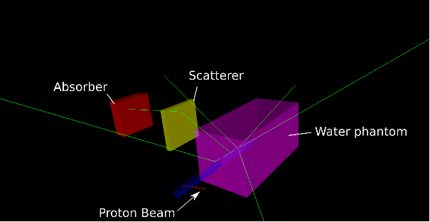
We are investigating Compton cameras to monitor particle therapy using simulations. Particle therapy is a kind of radiotherapy that destroy tumors using beams of protons or carbon nuclei. Compared to photon beams, particle beams show very useful properties when they interact with the patient: Very high energy doses can be precisely deposited in the tumor region, while organs at risks located behind the tumor are spared. On the other hand, the negative consequences of small errors in the treatment planing or changes in the patient are more severe than in radiotherapy. To avoid such problems, dedicated on-line verification is required for particle therapy . The development of verification methods is an active field of research. Verification methods exploit the radiation which emerges from the patient during the irradiation process. This radiation, which consists of different types of particles, is a natural consequence of the interactions between the therapeutic beam and the atoms of the patient. Several techniques have been identified as potentially useful, but there are still many aspects which require further investigation. In our project we are interested in prompt-gamma radiation. It consists of high-energy photons that can be registered with radiation detectors. Among such devices Compton cameras offer very promising features, such as the ability to produce images reflecting the origin of the prompt-gamma radiation. These images tell us indirectly where the particle beam deposited the maximum energy and stopped, thus providing a way to asses the irradiation.
Methods: To create an image from the data measured by the Compton camera, image reconstruction algorithms are needed. These algorithms act as a detective: Using the measured data as clues, and models of the physics as inspection tools, the algorithms can recreate the scenario which gave rise to the prompt-gamma radiation. As most Compton cameras are still prototypes under development, we use Monte-Carlo simulations to reproduce virtually what would occur in a real treatment scenario. Monte-Carlo simulations can produce very realistic data but require high computing power. Here we use the open-source simulation software GATE, which offers us a flexible framework to model different Compton cameras and therapeutic scenarios. Reconstruction algorithms rely on mathematical formulations and models of the radiation emission and detection processes. We use simulated data to test our reconstruction algorithms and the models as well as to provide feedback to the groups developing the Compton cameras with whom we collaborate. In this way, we can identify the challenges that Compton cameras will face when put into operation, and develop in advance strategies to cope with the problems. We also use simulations to test other uses of Compton cameras and prompt-gamma radiation [1,2]. For image reconstruction we have implemented several algorithms and physical models. The algorithms are iterative and, at this stage of research, require much computing power. Large amounts of data are handled, while several mathematical operations are carried out many times to create a reliable image. Next, we analyse the images to obtain useful information about the beam behaviour.
Results: We have successfully developed several image reconstruction procedures which have been adapted to three Compton cameras (currently under construction) [3,4]. Our approaches can significantly improve image quality and reduce the negative effects of statistical noise. Current limitations are the small size of the cameras, the complexity of the physical process behind the detection of high energy photons, and the high rates of prompt-gamma photons.
Ongoing work/outlook:We are developing approaches to compensate for the wrong information resulting from the high rates; we also investigate the best way to exploit some known a-priori in the reconstruction process. Our goal is to increase the accuracy of the verification procedure.
References
[1] A. Bolke, M. Zvolsky, N. Kohlhase, S. Seeger, M. Schaar and M. Rafecas, "Modelling of a Bi-Modal PET / Compton-Camera System for Non-Pure Positron Emitters," 2020 IEEE Nuclear Science Symposium and Medical Imaging Conference (NSS/MIC), Boston, MA, USA, 2020, pp. 1-3. doi:10.1109/NSS/MIC42677.2020.9507915
[2] J. Werner, N. Kohlhase, A. Bolke and M. Rafecas, "Neural networks for Compton camera imaging: Estimating initial and transferred energies of prompt gamma radiation". In: Student Conference Proceedings 2021. 10th Conference on Medical Engineering Science, 6th Conference on Medical Informatics, 4th Conference on Biomedical Engineering, 3rd Conference on Auditory Technology, 1st Conference on Biophysics, and 1st Conference on Robotics and Autonomous Systems. ISBN: 978-3- 945954-65-2.
[3] N. Kohlhase, T. Wegener, M. Schaar, A. Bolke, A. Etxebeste, D. Sarrut, and M. Rafecas, "Capability of MLEM and OE to Detect Range Shifts With a Compton Camera in Particle Therapy," in I EEE Transactions on Radiation and Plasma Medical Sciences, vol. 4, no. 2, pp. 233-242, 2020. doi: 10.1109/TRPMS.2019.2937675
[4] N. Kohlhase, M. Stille, A. Bolke, M. Zvolský and M. Rafecas, "Compton Camera Image Reconstruction with A-Priori Information from a Beam Tagging Hodoscope," 2020 IEEE Nuclear Science Symposium and Medical Imaging Conference (NSS/MIC), Boston, MA, USA, 2020, pp. 1-4. doi: 10.1109/NSS/MIC42677.2020.9507820
- Institute / Institutes:
- Institute of Medical Engineering (IMT)
- Affiliation:
- Universität zu Lübeck
- Image:
-